
Eye pressure tends to rise with age, and the risk of ocular hypertension increases after 40. According to data from the National Eye Institute, 4 to 10 percent of adults over age 40 in the United States have higher than normal eye pressure. If you are in this age group, regular screening is one of the simplest things you can do to protect your vision long term.
If a parent, sibling, or grandparent has been diagnosed with glaucoma, your risk of elevated eye pressure is higher. Genetics play a role in how your eye produces and drains fluid. Your eye doctor can factor your family history into a personalized monitoring plan.
The cornea is the clear front surface of your eye, like a windshield. If your cornea is thinner than average, standard pressure readings may actually underestimate the true pressure inside your eye. A simple measurement called pachymetry, which checks corneal thickness, helps your eye doctor get a more accurate picture of your risk.
High myopia (severe nearsightedness), a history of eye injury, or certain medical conditions like diabetes can also raise the chance of developing ocular hypertension. If any of these apply to you, your eye doctor may recommend more frequent pressure checks.
Your eye is not hollow. It is filled with a clear fluid called aqueous humor that nourishes the lens and cornea. Your eye produces this fluid all day long, and it drains out through a tiny mesh-like channel near the base of the iris, the colored part of your eye. Think of it like a sink with the faucet running nonstop. As long as the drain keeps up with the flow, pressure stays in a healthy range.
In ocular hypertension, the drain does not work as efficiently as it should. Fluid builds up faster than it can leave, and pressure inside the eye gradually rises. This is not something you feel. There is no pain, no redness, no sense of fullness. The only way to detect it is with a pressure measurement during an eye exam.
The optic nerve sits at the back of your eye and carries visual signals to your brain. When pressure inside the eye stays elevated over time, it can compress and damage the delicate nerve fibers. Once those fibers are lost, they do not grow back. This is how glaucoma develops. Ocular hypertension is not glaucoma, but it is the single biggest risk factor for it.
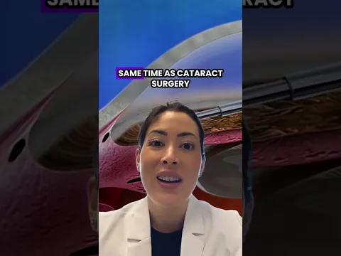
A landmark study published in JAMA Ophthalmology tracked patients with ocular hypertension over five years. The results, published in JAMA Ophthalmology, showed that only 4.4 percent of patients using pressure-lowering eye drops developed glaucoma, compared to 9.5 percent of untreated patients. That difference shows how much a simple daily drop can reduce your risk.
Elevated eye pressure raises your risk, but it does not mean glaucoma is inevitable. Many people live with slightly elevated pressure for years without any optic nerve damage. Your eye doctor looks at the full picture: your pressure readings, corneal thickness, optic nerve appearance, visual field results, and family history. All of these factors together help determine whether treatment makes sense for you right now or whether careful monitoring is the better path.

The most common test is tonometry, where your eye doctor uses a small instrument to measure the pressure inside your eye. You may feel a brief, gentle puff of air or a light touch on the surface of your eye. The test takes just a few seconds and gives your doctor a clear number to work with. Normal eye pressure typically falls between 10 and 21 millimeters of mercury.
Your eye doctor will look at the optic nerve directly using specialized imaging. This shows whether the nerve head looks healthy or whether there are early signs of thinning or cupping, which is when the center of the nerve starts to hollow out. Advanced imaging technology can detect changes so subtle that they would be invisible during a standard exam.
A visual field test maps your peripheral (side) vision. Glaucoma typically affects side vision first, and this test can pick up blind spots before you ever notice them. If your pressure is elevated, your eye doctor may run a visual field test as a baseline and repeat it over time to watch for any changes.
Because corneal thickness affects how pressure readings are interpreted, your eye doctor may measure it with a painless test called pachymetry. Thinner corneas can make pressure appear lower than it truly is. This one measurement can change your entire risk profile.
For most patients with ocular hypertension, daily prescription eye drops are the first step. These drops either reduce the amount of fluid your eye produces or help it drain more efficiently. They are painless to use and highly effective at bringing pressure into a safer range. Your eye doctor will check your pressure at follow-up visits to make sure the drops are working and adjust the type or dosage if needed.
If eye drops are not enough on their own, or if you have trouble using them consistently, your eye doctor may refer you for a gentle laser procedure called selective laser trabeculoplasty. This treatment uses focused light to open up the drainage channels in your eye, helping fluid flow out more freely. The procedure is performed on an outpatient basis and typically takes just a few minutes. Our eye doctors at Insight Vision Optometry, Las Vegas co-manage this process, handling your evaluation, referral, and follow-up care.
Not every case of elevated eye pressure requires immediate treatment. If your pressure is only mildly elevated and your optic nerve looks healthy, your eye doctor may recommend a 'watch and wait' approach with regular checkups every few months. This is not ignoring the problem. It is a deliberate, evidence-based strategy that avoids unnecessary medication while keeping a close eye on any changes.
During a comprehensive eye exam, your eye doctor will measure your intraocular pressure, examine the optic nerve, and review your overall eye health. If your pressure comes back above the normal range, they will discuss what that number means and whether additional testing is needed. This conversation is straightforward and pressure-free. You will understand exactly where you stand before you leave.
If your eye doctor finds elevated pressure, they will likely schedule a follow-up visit to recheck the measurement. Eye pressure can fluctuate throughout the day, so a single high reading does not mean you have ocular hypertension. Two or more elevated readings, combined with other risk factors, give a clearer picture. Over time, your eye doctor builds a pressure history that shows trends and helps guide treatment decisions.
Managing elevated eye pressure is not dramatic. If you need drops, they become part of your daily routine like brushing your teeth. If you are being monitored, you come in for checkups on a regular schedule. Most patients continue their normal activities without restrictions. The key is consistency: staying on track with your drops or your monitoring visits is what keeps your optic nerve protected year after year.
Your visit begins with a thorough evaluation, including tonometry, optic nerve imaging, and corneal thickness measurement. Our eye doctors in Spring Valley use advanced diagnostic technology to get the full picture of your eye health in a single appointment.
After testing, your eye doctor will sit down with you and explain what the numbers mean. They will walk you through your individual risk factors, including age, family history, corneal thickness, and optic nerve appearance. Together, you will decide whether to start treatment or begin a monitoring schedule.
If treatment is recommended, your eye doctor will prescribe the appropriate drops and show you how to use them properly. If monitoring is the right choice, they will schedule follow-up visits at the right intervals. Either way, you will leave with a clear plan.
Ocular hypertension is a long-term condition that benefits from long-term attention. Insight Vision Optometry, Las Vegas supports patients in Southwest Las Vegas and the surrounding community with consistent follow-up care, pressure tracking over time, and adjustments to your plan whenever the data suggests a change.


