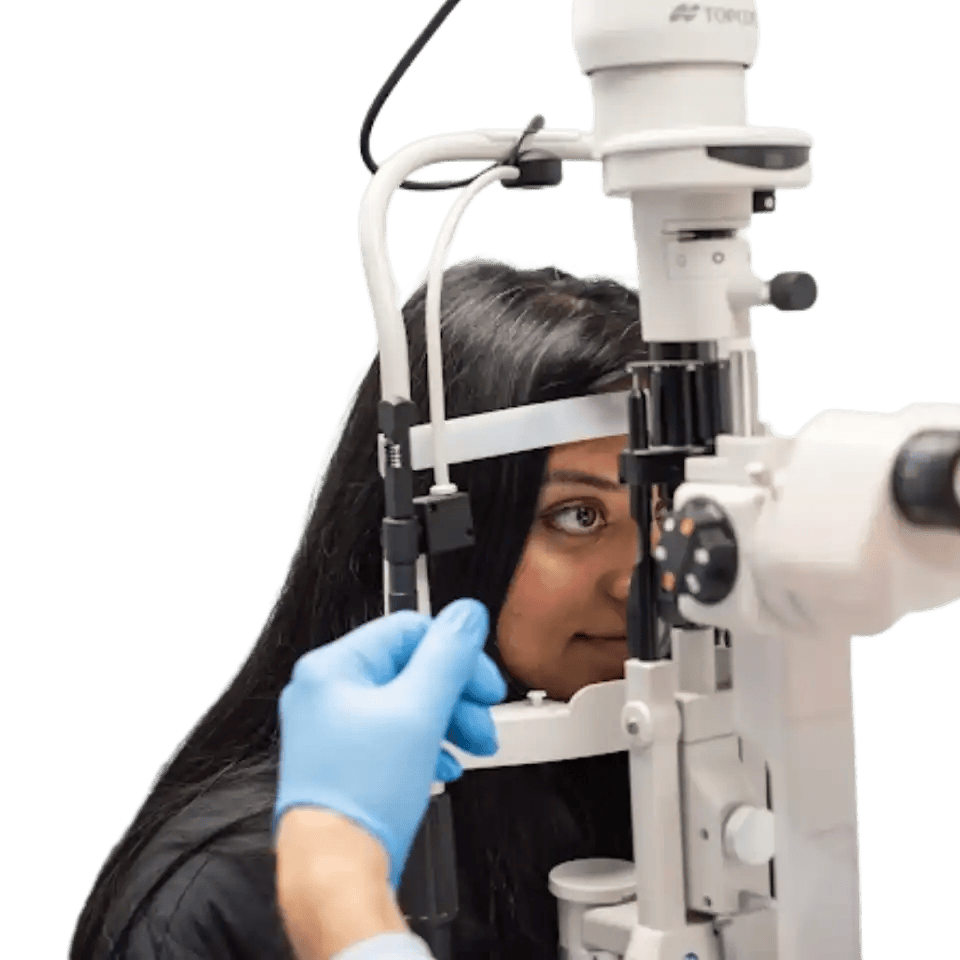
There is no single monitoring schedule that fits every keratoconus patient. Your age, stage of disease, and individual risk factors all shape how often your corneas need to be imaged. Our doctors tailor each patient's schedule to their specific situation.
If your keratoconus has been stable over multiple visits, corneal imaging is typically scheduled every 6 to 12 months. Each appointment compares your current corneal maps to your previous scans, looking for any shift in steepness, thickness, or shape. Even when everything looks stable, these appointments matter because late progression, while uncommon, does happen and is best caught early.
Young patients with keratoconus typically need corneal imaging every 3 to 6 months. Pediatric keratoconus tends to progress more rapidly than in adults, and in many cases it presents at a more advanced stage at the time of diagnosis. Parents should prioritize these appointments even when their child's vision seems unchanged, because the cornea can be shifting before any vision changes are noticeable.
Certain factors place patients in a higher-risk category that may call for closer monitoring. Your doctor will review your imaging history and personal health profile to determine the right interval for you.
If any of these apply to you, your doctor may schedule imaging more frequently than the standard 6-to-12-month window.
Receiving treatment for keratoconus does not end the need for monitoring. In fact, the period after treatment is one of the most important times to keep a close watch on your corneas. Our doctors continue imaging after every type of intervention to confirm your results and catch any changes early.
Corneal cross-linking is a procedure that uses UV light and riboflavin drops to strengthen the cornea and slow or halt progression. However, cross-linking does not always provide a permanent halt, particularly in younger patients. Research has shown that a meaningful portion of pediatric patients can resume corneal steepening within a few years of the procedure. Because of this, at least five years of follow-up imaging is recommended after cross-linking, with annual or semi-annual scans continuing throughout.
Patients who have had intrastromal corneal ring segments, keratoplasty (corneal transplant), or other surgical interventions require monitoring of both the treatment results and the underlying cornea. Post-surgical imaging tracks graft health, ring position, and any corneal changes occurring outside the treated area. Monitoring visits are typically more frequent in the first year after surgery and then taper as the cornea stabilizes.
Keratoconus often slows significantly or stabilizes on its own by the mid-thirties to forties, as the cornea naturally becomes stiffer with age. Even so, documented cases of progression in patients over age 48 mean that monitoring is a lifelong commitment. At minimum, annual corneal imaging is recommended even after many years of confirmed stability, because late changes can still occur and are best caught before they affect your vision or your contact lens fit.

A keratoconus monitoring visit is different from a standard eye exam. It is built around precise corneal imaging and careful comparison with your past scans. Understanding what to expect can help you make the most of each appointment.
Every monitoring visit includes corneal topography or tomography, which creates a detailed map of your cornea's shape, curvature, and thickness. Your doctor also performs a vision check with your current correction to confirm your prescription is still working well. Depending on your situation, additional testing may include OCT epithelial mapping or corneal biomechanical assessment to evaluate the structural integrity of the cornea.
Your doctor compares the new scans directly to your baseline and all previous imaging to identify trends over time. The key measurements reviewed at each visit include the following.
Your doctor uses all of these data points together to judge whether your condition is holding steady or beginning to shift.
If your scans are stable visit after visit, your doctor continues the current schedule with no changes needed. If progression is detected, your doctor will discuss intervention options, such as cross-linking, and help you understand what the findings mean for your vision and your contact lens correction. If your vision with your current correction has declined, a lens change or other adjustment may be recommended. The results of every monitoring visit directly guide the next step in your care.

The most accurate picture of your keratoconus comes from consistent, uninterrupted imaging over time. There are practical steps you can take between and during appointments to support the quality of your monitoring and strengthen the partnership with your doctor.
Corneal changes in keratoconus can be subclinical, meaning the imaging shows progression before you notice any difference in your daily vision. Skipping even one visit creates a gap in your imaging history that makes it harder to detect trends accurately. Catching progression while it is still subtle gives you and your doctor the most options for intervention. Treat every scheduled imaging appointment as a necessary part of maintaining your vision health.
Pay attention to any changes in your vision between appointments, particularly increased blur, more glare, or halos around lights at night. Note whether your glasses or contact lenses seem less effective than they did after your last fitting. If these changes are sudden or significant, contact your doctor's office rather than waiting for your next scheduled visit. Your observations add important context to the objective measurements your doctor takes at each appointment.
If you relocate or transfer care to a new provider, bring your previous corneal imaging files with you. Serial comparison of maps taken on consistent equipment gives the most accurate picture of your progression over time. Ask your current doctor for copies of your imaging data before any planned transition. A continuous imaging history is one of the most valuable tools in managing keratoconus effectively.
These questions address common concerns patients have about fitting monitoring into their lives and understanding what their results mean.
Many insurance plans do cover corneal imaging when keratoconus is an active diagnosis, so it is worth confirming your benefits before assuming you will face a large out-of-pocket cost. If coverage is limited, talk with our team about payment plans or financing options that can make regular visits more manageable. Delaying or skipping monitoring to avoid cost often leads to larger and more expensive problems down the road if progression goes undetected.
Home vision checks, such as covering one eye and looking at a straight-edged object to detect distortion, can give you a rough sense of whether something feels different. These informal checks are not a substitute for clinical imaging but can prompt you to call your doctor if something seems off. If you notice a sudden change in distortion, blur, or clarity, that warrants a call to our office rather than waiting for a scheduled appointment.
The corneal imaging schedule is determined by your keratoconus status, not by the type of contact lens you wear. However, your doctor will also check that your current lenses are still fitting correctly at monitoring visits, since a changing cornea can affect lens fit, comfort, and vision performance even when you feel fine. Scleral lens wearers in particular benefit from having fit and corneal imaging reviewed together.
A focused monitoring visit typically takes between 20 and 45 minutes depending on which tests are included. If your doctor adds biomechanical testing or OCT epithelial mapping, the visit may run slightly longer. We recommend building in a little extra time so results can be reviewed with you before you leave.
A gap in your monitoring history does not mean care cannot continue, but it does mean your doctor will need to establish a new baseline before trends can be accurately assessed. Bring any previous imaging files you have access to, even if they are older. From there, more frequent visits may be recommended for a period to determine whether your cornea is currently stable or actively changing.
There is no age or stage at which monitoring can be safely stopped altogether. The frequency may decrease as you accumulate years of confirmed stability, particularly in adulthood, but annual imaging remains the standard recommendation for life. If you are unsure whether your current schedule is appropriate, bring that question directly to your doctor so the interval can be reviewed based on your full imaging history.
At Insight Vision Optometry, Las Vegas, our doctors bring specialized expertise in keratoconus management and corneal imaging to every monitoring visit. We use advanced topography and tomography technology to track even subtle changes with precision, giving you and your doctor the clearest possible picture of your corneal health. Whether you are newly diagnosed, post-treatment, or managing long-term stability, we are here to keep your vision protected at every stage. Contact our office to schedule your next monitoring appointment and experience the level of care that sets us apart.


