
Corneal dystrophy and corneal degeneration both affect the clarity and function of the cornea, but they develop in very different ways. Identifying which condition is present shapes every decision that follows, from how closely your vision is monitored to which treatments are most appropriate.
The cornea is a clear, dome-shaped tissue that sits at the very front of your eye. It bends incoming light so that it can land precisely on the retina at the back of the eye, making sharp vision possible. The cornea also acts as a protective barrier against dust, debris, and harmful microorganisms. It is made up of five distinct layers, and each layer plays a specific role in maintaining transparency and structural strength.
Corneal dystrophy is a hereditary condition, meaning it is passed down through families through a change in a specific gene. That gene change causes abnormal deposits to build up within one or more layers of the cornea over time. Because dystrophy is genetic, it almost always affects both eyes, though sometimes one eye may show more noticeable changes earlier. Many forms follow what is called an autosomal dominant inheritance pattern, meaning that one parent who carries the gene has a chance of passing it to each child.
Corneal degeneration is acquired rather than inherited. It can develop as a result of aging, prolonged ultraviolet light exposure, chronic inflammation, or other systemic health conditions. These changes often begin at the outer edge of the cornea and may affect one eye more noticeably than the other. Common examples include arcus senilis (a grayish ring at the corneal edge), band keratopathy (calcium deposits across the cornea), and Salzmann nodular degeneration (raised, grayish spots on the corneal surface).
Eye doctors use a system called IC3D to organize corneal dystrophies by which layer of the cornea is affected. The four main categories are epithelial, epithelial-stromal, stromal, and endothelial. Knowing which layer is involved helps guide both the diagnosis and the most appropriate treatment plan. This classification system is now used internationally to bring consistency to how these conditions are described and managed.
Symptoms of corneal dystrophy and degeneration often begin subtly and become more noticeable over months or years. Some people notice changes in how they see light or how their vision feels at different times of day. Recognizing these early signs and bringing them to the attention of your eye doctor gives you the widest range of care options.
Blurred or hazy vision is often the first sign that something is affecting the cornea. It may come and go at first, a bit like looking through a fogged-up window. Reading small print, seeing clearly at a distance, or recognizing faces across a room may all become harder. In Fuchs endothelial dystrophy (a condition affecting the innermost layer of the cornea), vision is often worst in the morning and gradually improves as the day goes on and fluid naturally evaporates from the corneal surface.
Many people with corneal conditions describe a feeling of sand or grit stuck beneath the eyelid, even when nothing is there. Sharp pain on waking is a particularly telling symptom that may point to a recurrent corneal erosion, a condition where the outermost layer of the cornea repeatedly lifts away from the layer beneath it. This pattern is closely associated with epithelial basement membrane dystrophy and can range from mildly uncomfortable to acutely painful.
Increased sensitivity to light, known as photophobia, is a common complaint with many corneal conditions. Bright sunlight, overhead lighting, and oncoming headlights at night can all feel unusually harsh or painful. Glare and starbursts around lights are especially common at night, which can make driving after dark a challenge. Many people find themselves reaching for sunglasses more often or avoiding brightly lit environments.
The eye sometimes produces excess tears as a response to surface stress or irritation. Some people wake to find thick, stringy mucus near the eyes. Dry eye symptoms can overlap with corneal conditions because the tear film, the thin protective layer of moisture across the cornea, may not function well when the corneal surface is irregular or damaged. Managing both the corneal condition and tear film quality is often part of a complete care plan.
Fluctuating vision throughout the day is a useful clue when your doctor is trying to identify the cause of your symptoms. In Fuchs dystrophy, morning haziness often fades as the day progresses because excess fluid gradually evaporates from the cornea. In conditions related to dry eye or ocular surface disease, vision may worsen as the day goes on. Keeping a simple note about when your vision feels clearest and when it is most difficult can be genuinely helpful at your appointment.

Some causes of corneal conditions are beyond your control, such as inherited genes or the natural process of aging. Others can be influenced by lifestyle choices and how closely you manage your overall health. Understanding these factors helps you and your eye doctor make informed decisions about monitoring and prevention.
All corneal dystrophies trace back to changes in specific genes that affect how the cornea builds, maintains, and repairs itself. Many dystrophies follow an autosomal dominant inheritance pattern, meaning that one copy of the altered gene is enough to cause the condition. Others require two copies, one from each parent, before the condition develops. Genetic counseling is available for families who want to understand their risk and explore testing options for relatives who may not yet show symptoms.
The corneal cells that keep the surface clear and smooth become less efficient at renewal as we age. Decades of exposure to ultraviolet light, wind, dust, and environmental irritants can accelerate that process. Chronic allergies and long-term inflammation also contribute to corneal surface changes over time. Fuchs endothelial dystrophy, for example, most often becomes symptomatic after age 50, even though the genetic tendency has been present from birth.
Women develop Fuchs endothelial dystrophy at a significantly higher rate than men, with some estimates suggesting it occurs two to three times more often in women. The exact reason for this difference is not fully understood, and research into possible hormonal and genetic connections is ongoing. This pattern means that women with a family history of Fuchs dystrophy may benefit from earlier and more regular corneal monitoring.
Certain systemic health conditions affect the cornea in indirect but meaningful ways. Diabetes can reduce corneal sensation and slow the healing of surface injuries. Autoimmune conditions such as rheumatoid arthritis can cause severe dry eye and contribute to corneal surface damage over time. Some medications, including certain antihistamines, reduce natural tear production, which can worsen a cornea that is already under stress. Managing these underlying conditions is an important part of corneal care.
A corneal scratch, a chemical splash, or a burn from a welding arc or UV lamp can all leave lasting structural changes to the corneal layers. Previous eye surgeries, including cataract removal and laser vision correction, can also influence how the cornea responds over time. For people who already have Fuchs dystrophy, cataract surgery carries additional considerations, since the procedure may accelerate endothelial cell loss if pre-operative planning does not account for the existing condition.
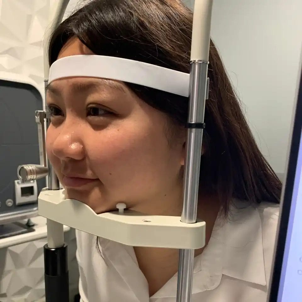
Diagnosis relies on a combination of your personal and family history, a detailed clinical examination, and precise imaging of the corneal layers. Several tests are often used together because no single measurement tells the whole story. The goal is to identify which layer is affected, how much change has already occurred, and which direction the condition is likely to move over time.
Every corneal evaluation begins with a careful conversation about your symptoms, how long they have been present, and whether any family members have had similar problems. The slit lamp is a specialized microscope paired with a focused beam of light that allows your doctor to examine each corneal layer in magnified detail. Deposits, surface irregularities, areas of swelling, and early structural changes all become visible under the slit lamp that would be invisible to the naked eye.
Corneal topography creates a detailed color-coded map of the curvature of the entire corneal surface. This test is particularly useful for identifying irregular shapes, steep zones, or thinning patterns that can indicate keratoconus or other structural changes. Pachymetry measures the thickness of the cornea at multiple points. Thickness readings are used to monitor swelling in Fuchs dystrophy and to assess corneal health before and after surgical procedures.
A specialized test called specular microscopy photographs and counts the endothelial cells, the innermost layer of pump cells that keep the cornea clear by removing excess fluid. Loss of these cells is a defining feature of Fuchs endothelial dystrophy. Tracking cell density and cell shape over time helps your doctor assess how quickly the condition is progressing and whether surgical planning should begin. These cells do not naturally regenerate, which is why monitoring their count is so important.
For suspected corneal dystrophy, genetic testing can identify the specific mutation responsible and clarify the inheritance pattern for the family. Even without formal laboratory testing, a thorough review of family history combined with clinical exam findings in the patient and, when possible, in relatives can provide useful diagnostic information. Results from genetic testing can also inform how frequently examinations should be scheduled and help identify relatives who may benefit from early evaluation.
Treatment is tailored to the specific condition, the layer of the cornea involved, and the degree to which vision or comfort is affected. Many people manage their condition effectively with drops and specialty contact lenses for years. When those options are no longer sufficient, modern surgical techniques offer excellent outcomes with faster recovery than was possible in the past.
Artificial tears are a foundational part of corneal care and help keep the surface properly moisturized, reducing friction and irritation. Hypertonic saline drops and ointments work by drawing excess fluid out of the cornea, which is particularly useful for managing morning haziness in Fuchs dystrophy. Antibiotic drops may be prescribed to protect the eye during healing after a corneal erosion. Following a consistent drop regimen as directed by your doctor can meaningfully improve daily comfort and visual quality.
Specialty contact lenses play an important role in managing both corneal dystrophy and degeneration. A therapeutic soft bandage lens can cover and protect the corneal surface while a recurrent erosion heals. Scleral lenses are large-diameter rigid lenses that vault completely over the cornea, creating a smooth, fluid-filled optical surface that can dramatically improve vision in irregular or damaged corneas. Rigid gas permeable lenses are another option for sharpening vision when the corneal surface is too uneven for standard glasses or soft contacts to work well.
Several minimally invasive procedures can provide meaningful relief without surgery. Punctal plugs are tiny devices placed in the tear drainage channels to help retain your natural tears on the eye's surface for longer. Phototherapeutic keratectomy, called PTK, uses a precise laser to smooth rough or irregular corneal tissue and can also remove shallow corneal deposits that are affecting vision. Corneal cross-linking is a procedure that uses UV light and riboflavin eye drops to strengthen corneal tissue, and it can help slow or halt the progression of keratoconus and related conditions.
For patients with Fuchs endothelial dystrophy who are no longer managing well with drops and lenses, a surgical procedure called DMEK (Descemet membrane endothelial keratoplasty) has become the preferred surgical treatment. DMEK involves transplanting only the thin innermost layer of a donor cornea rather than replacing the entire cornea. This targeted approach results in faster recovery, a lower risk of rejection compared to older transplant techniques, and vision outcomes that often reach 20/25 or better in a substantial portion of patients. Our doctors work closely with experienced corneal surgeons to coordinate care when surgical referral is appropriate.
A full-thickness corneal transplant, known as penetrating keratoplasty, may still be necessary when disease involves the deeper stromal layers and a partial-thickness technique is not sufficient. For carefully selected patients with early-to-moderate Fuchs dystrophy who still have healthy endothelial cells at the outer edge of the cornea, a newer approach called Descemet stripping only (DSO) removes the damaged central endothelium without a donor graft, allowing the patient's own peripheral cells to migrate inward and restore clarity. DSO is appropriate only in specific cases, and your doctor will discuss whether it is a realistic option during your evaluation.
These questions address some of the practical decisions and concerns that come up when living with a corneal condition. If your situation raises questions not covered here, bring them to your next appointment.
Total blindness from these conditions is rare, particularly with consistent monitoring and timely treatment. Most people retain functional vision throughout their lives with appropriate care. When vision does decline significantly, corneal transplant surgery has a strong track record of restoring clarity. The key factor is staying engaged with regular follow-up so that any meaningful change is caught early enough to act on it.
Protective habits make a genuine difference over the long term. Wearing wrap-style UV-blocking sunglasses whenever you are outdoors reduces cumulative UV damage that can accelerate surface changes. Avoiding rubbing your eyes is especially important because repeated mechanical friction can worsen many corneal conditions. Other helpful habits include wearing safety glasses during yard work or sports, keeping blood sugar and allergies well managed, and following all contact lens care instructions your doctor provides. None of these steps replace professional monitoring, but they can reduce the burden on the corneal surface over time.
Cataract surgery is absolutely possible for patients with Fuchs dystrophy, but it requires careful advance planning. Your surgeon will measure endothelial cell density before the procedure to understand how much reserve the cornea has. Special surgical techniques that minimize stress to the pump cells are used, and your surgical team may recommend a combined procedure that addresses both the cataract and the corneal disease at the same time using DMEK. Open communication between your optometrist and your cataract surgeon before the procedure is essential for a safe outcome.
Children from families with a known corneal dystrophy should be examined by an eye doctor experienced with corneal conditions, even if they currently have no symptoms. Many dystrophies can be detected on clinical examination before they cause any vision problems, and establishing a baseline early allows for meaningful comparison over time. Genetic counseling can also clarify the probability of inheritance based on the specific condition in the family. Early detection does not always mean early treatment, but it ensures that no changes are missed during critical developmental years.
Yes, and in many cases waiting is the right approach. Transplant surgery is typically considered when vision has declined to a degree that interferes with driving, reading, working, or other important daily activities, and when non-surgical options are no longer providing adequate relief. Many patients manage comfortably with drops, specialty lenses, and minor procedures for years before surgery becomes necessary. Your doctor will track changes at each visit and have an honest conversation with you about when the balance between benefits and risks of surgery begins to favor proceeding.
Some symptoms warrant same-day contact with your eye doctor rather than waiting for a scheduled appointment. Sudden sharp pain on waking, a new significant drop in vision, a red eye with discharge, or any sensation of something lodged in the eye that does not resolve should all be evaluated promptly. New or worsening halos around lights, especially if they appear quickly rather than developing gradually, also deserve urgent attention. These signs can indicate an acute erosion, an active infection, or a rapid increase in corneal swelling, all of which respond better to treatment when addressed quickly.
Our team at Insight Vision Optometry brings specialized training in corneal conditions, advanced imaging technology, and the experience of managing complex cases from across Nevada and beyond. Whether you are newly diagnosed, monitoring a known condition, or simply want a thorough evaluation, we are here to give you answers and a plan you can feel confident in. We welcome you to schedule your corneal health visit with us in Las Vegas.


