The cornea is the clear dome at the very front of the eye. It depends on a healthy tear film and a well-functioning immune system to stay transparent, smooth, and strong. Autoimmune diseases disrupt both, often in multiple ways at once.
Autoimmune disease causes the body's immune system to mistakenly attack its own tissue. In the eye, this can take several forms depending on which condition is involved and how well it is controlled.
Each of these changes can progress quietly, which is why routine eye exams are so important for anyone with an autoimmune diagnosis.
The cornea gets most of its oxygen directly from the air and most of its nutrients from the tear film. When autoimmune disease attacks the glands that produce tears, the corneal surface loses its main source of protection. A dry surface becomes fragile, breaks down more easily, heals slowly, and is more open to infection. In some conditions, the immune system sends destructive cells directly to the cornea itself, causing inflammation and tissue damage that goes beyond simple dryness.
Several autoimmune conditions are known to involve the eye, particularly the cornea. Sjogren syndrome is one of the most common causes of severe dry eye. Rheumatoid arthritis can cause a dangerous form of corneal thinning near the outer edge. Lupus leads to surface dryness and light sensitivity. Graves disease, which affects the thyroid gland, can push the eye forward and expose the cornea to air even during sleep. Inflammatory bowel diseases such as Crohn disease and ulcerative colitis can also cause eye inflammation, including corneal involvement, especially during flares.
Not everyone with an autoimmune condition will develop corneal disease, but certain factors increase the risk. Women are more likely than men to develop many autoimmune conditions, and tear production naturally decreases with age, which compounds the dryness. People with long-standing or poorly controlled disease, those taking medications with drying side effects, and those with a history of eye surgery all face a higher baseline risk.
Different autoimmune diseases affect the cornea in different ways. Understanding how each condition behaves helps our team tailor your care plan and helps you recognize warning signs early.
Sjogren syndrome directly attacks the glands that produce tears and saliva, making it one of the most common causes of severe dry eye in clinical practice. Patients often describe a sandy or gritty feeling, burning, and vision that blurs by mid-afternoon. Without consistent treatment, the cornea can develop surface breaks, small scars, and persistent sores. With the right combination of drops, prescription medications, and supportive care, most patients can maintain a healthy corneal surface and preserve their vision.
Rheumatoid arthritis is known for joint pain, but it can also cause a serious corneal complication called peripheral ulcerative keratitis, which is thinning and breakdown of the cornea near its outer edge. This condition is uncommon but carries real risk of corneal perforation if not caught and treated early. Patients with poorly managed rheumatoid arthritis face the greatest risk. Keeping joint disease well controlled through medication often provides meaningful protection to the eyes as well.
Systemic lupus erythematosus can cause dryness, light sensitivity, and surface changes on the cornea. Some patients also notice burning or discomfort that is not explained by the level of visible dryness. A thorough eye exam that evaluates the tear film, the corneal surface, and the inner eye is recommended at diagnosis and at regular intervals. Most lupus patients respond well to artificial tears and close monitoring, though some require prescription drops or additional treatment.
Graves disease causes the thyroid gland to become overactive and can cause the tissue behind the eyes to swell, pushing the eyes forward. This forward displacement, combined with eyelids that may not fully close, leaves the cornea exposed to air during sleep and throughout the day. Without protection, the surface dries rapidly and can develop painful sores. Nighttime ointments, moisture-retaining eyewear, and humidified sleep environments are first-line protective measures. In more severe cases, surgical intervention is necessary to reduce exposure and protect the cornea.
Crohn disease and ulcerative colitis are primarily gut conditions, but both can involve the eyes during active flares. Corneal inflammation in this setting is less common than with other autoimmune diseases, but it can develop quickly with redness, pain, and vision changes. Coordinated care between your gastroenterologist and your eye doctor is especially important, since controlling the underlying bowel disease often helps resolve the eye involvement at the same time.

Many corneal changes in autoimmune disease start with mild symptoms that are easy to dismiss. Knowing which signs are urgent and which can wait for a scheduled visit helps you act at the right moment.
Early eye involvement in autoimmune disease is often subtle. You may notice a gritty or burning feeling, mild light sensitivity, or vision that seems clearer after blinking. Tear output may actually drop while the eye feels watery, because the surface triggers reflex tearing in response to dryness. These mild signs are worth noting and raising at your next eye visit, even if they seem minor.
Some eye symptoms should not wait. Sharp eye pain, sudden vision loss, a red eye with a white or gray spot on the cornea, or severe sensitivity to light all require same-day evaluation. A cornea weakened by autoimmune disease can break open quickly under the right conditions, and early intervention can prevent permanent vision loss. If you have a known autoimmune condition and develop sudden eye redness with pain, seek care the same day.
Just as joints can flare with rheumatoid arthritis or rashes can return with lupus, the eyes can flare with autoimmune disease. New halos around lights, a sudden increase in light sensitivity, or a red eye that appears alongside body-wide symptoms may all mean the disease is becoming more active. Eye signs sometimes appear before other body signs, which can give your broader medical team an early signal to adjust treatment.

Diagnosing corneal disease in the setting of autoimmune conditions requires a thorough, layered approach. Our doctors combine detailed history-taking, specialized testing, and advanced imaging to build a complete picture of your corneal health.
A complete medication list is one of the most helpful things you can bring. Include all prescription drugs, over-the-counter medications, vitamins, and herbal supplements. Note your autoimmune diagnosis, when it was confirmed, and any recent flares or hospitalizations. If you have had recent blood work, bring a copy. The more context your eye doctor has, the more precisely your care can be tailored.
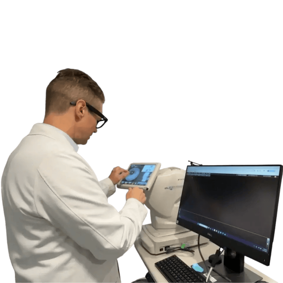
Our doctors use a slit lamp microscope to examine each layer of the cornea in detail. A dye called fluorescein is applied to the eye surface to reveal any areas of breakdown that may not be visible otherwise. A small paper strip placed briefly under the eyelid measures how much tear the eye produces. A separate dye test measures how quickly the tear film breaks apart on the cornea, which reflects both tear quality and gland health.
For patients with suspected corneal thinning or structural change, additional tests are valuable. Pachymetry measures the exact thickness of the cornea. Corneal topography produces a detailed map of the corneal surface shape, which helps detect subtle thinning or irregularity early. Anterior segment optical coherence tomography, known as AS-OCT, provides a cross-sectional view of the corneal layers, allowing our team to track changes over time with high precision.
Your eye doctor, rheumatologist, and primary care physician each see one part of the full picture. Our doctors work directly with your medical team to make sure that drug changes, disease flares, and new findings are shared across offices. When everyone is informed, treatment decisions are safer and more effective. If you have a rheumatologist managing your autoimmune condition, letting both teams communicate is one of the most protective steps you can take for your eyes.
Treatment is always matched to how your cornea looks on examination and how the disease is behaving. Our doctors, including Dr. Stephanie Woo, Dr. Nathan Schramm, and Dr. Thanh Mai, bring advanced corneal and specialty lens expertise to building individualized plans for each patient.
Most treatment plans begin with preservative-free artificial tears used consistently throughout the day. A gel or ointment at bedtime adds moisture protection through the night, when the eyes may not fully close. A humidifier in the bedroom, wraparound glasses when outdoors in wind or dry air, and warm compresses on the lids to unblock the oil-producing meibomian glands all support the tear film. These steps are simple, but their consistent use makes a significant difference over time.
When over-the-counter drops are not enough, prescription options are added. Cyclosporine and lifitegrast are anti-inflammatory drops that reduce immune activity in the tear glands, helping them produce better-quality tears. Short courses of steroid drops can calm an acute flare, though long-term steroid use carries risks including elevated eye pressure and cataract formation. Small plugs placed into the tear drainage openings, called punctal plugs, help preserve natural tears on the eye surface for longer periods.
When corneal tissue is at serious risk of breakdown or the surface has not responded to standard care, more advanced interventions are considered. An amniotic membrane, a thin biological tissue, can be placed on the cornea to promote healing and reduce inflammation. A soft bandage contact lens can shield a fragile surface from the mechanical irritation of blinking. In severe exposure cases, a partial tarsorrhaphy, a procedure in which the eyelids are partially closed with sutures, protects the cornea from persistent air exposure.
For patients with advanced dryness or an irregular corneal surface, scleral contact lenses offer significant relief. These are large-diameter rigid lenses that vault entirely over the cornea and rest on the white of the eye, creating a fluid reservoir that keeps the corneal surface continuously bathed in a moisture-rich environment throughout the day. Dr. Woo, Dr. Schramm, and Dr. Mai have deep expertise in fitting scleral lenses for patients with complex ocular surface disease. Autologous serum drops, which are drops made from the patient's own blood and contain natural growth factors, can also support healing in difficult cases.
Medications you take for your autoimmune condition play a direct role in corneal health. Better control of the underlying disease generally means less inflammation reaching the eye. In select cases, our doctors may communicate with your rheumatologist about whether a medication adjustment could reduce corneal risk. This is always a shared decision involving all treating physicians, with the goal of protecting both the joints and the eyes at the same time.
Day-to-day habits and consistent follow-up care are central to protecting your corneas over the long term. Small, repeatable actions add up to meaningful protection when done consistently.
Staying well hydrated supports tear production throughout the day. Foods rich in omega-3 fatty acids, including fish, flaxseed, and walnuts, may support the oil layer of the tear film. Taking screen breaks every twenty minutes reduces surface evaporation during computer work. Using drops on a scheduled routine rather than waiting until your eyes feel dry is more effective, since dryness felt on the surface often means the surface is already stressed.
The right visit frequency depends on how active your disease is and how the cornea looks on examination. Most patients with mild, well-controlled autoimmune disease are seen once a year. Patients with Sjogren syndrome, active rheumatoid arthritis, or a history of corneal thinning benefit from visits every three to six months. After any disease flare, new medication, or drug change, scheduling a follow-up within a few weeks is a good general guideline. Any sudden change in vision or comfort should prompt a same-day call.
A simple log of eye symptoms, drop use, and any medication changes is one of the most practical tools you can bring to each appointment. Sharing this information with both your eye doctor and your rheumatologist helps both teams spot patterns and respond before small changes become serious problems. Many patients find that keeping a brief weekly note on their phone makes this easy to maintain over time.
These answers address specific questions our patients commonly raise about autoimmune disease, corneal health, and how to navigate care across multiple providers.
Most people with autoimmune conditions maintain good vision throughout their lives, particularly when corneal disease is identified early and managed consistently. The risk of serious vision loss is real but significantly reduced by routine examinations, good systemic disease control, and prompt treatment of new findings. The patients who face the most serious outcomes are generally those who delayed care or had disease that was difficult to control for an extended period. Our team works to reduce that risk through proactive monitoring and individualized planning.
Yes, absolutely. Even well-controlled systemic disease can cause quiet corneal changes that are only visible on a slit lamp examination. Blood tests and joint markers do not reflect what is happening at the corneal surface. Routine eye exams serve as a complementary surveillance tool and give your full medical team information that blood work alone cannot provide. They are one of the most practical steps you can take to protect long-term vision.
It depends on the severity of your dryness and the condition of your corneal surface. Mild dry eye often still allows for daily soft lens wear with appropriate drops and a shorter daily wear schedule. More advanced dryness can make standard contact lenses uncomfortable and increase surface irritation. Scleral lenses are a well-established option for many autoimmune patients because they hold a reservoir of fluid against the cornea rather than sitting on the dry surface. Our doctors can evaluate your specific situation and recommend a contact lens plan that is safe for your eyes.
Do not stop or reduce any medication without speaking to the prescribing doctor first. Many medications used to manage autoimmune conditions have drying effects on the eye, but the risks of stopping or reducing those medications are generally much greater than the eye side effects, which can usually be managed with additional drops or other treatments. If your eye discomfort is significant, let your eye doctor and rheumatologist know together so they can make a coordinated decision about whether any adjustment is warranted.
Standard soft contact lenses sit directly on the corneal surface and can draw moisture away from an already dry eye. Scleral lenses, by contrast, vault entirely over the cornea and create a sealed liquid chamber between the lens and the eye. This chamber continuously bathes the corneal surface in a saline solution throughout the day, providing a level of moisture that drops alone often cannot match. For patients with Sjogren syndrome or other severe dry eye conditions, scleral lenses can restore comfort and vision quality that other treatments have not achieved.
Diet alone cannot replace medical treatment, but certain nutritional choices may support the tear film. Omega-3 fatty acids found in oily fish, flaxseed, and walnuts have been studied for their potential role in supporting the meibomian gland oil layer that stabilizes tears. Staying well hydrated throughout the day supports tear production in general. Reducing caffeine and alcohol intake may also help, as both have dehydrating effects. If your autoimmune condition affects how you digest or absorb nutrients, working with a registered dietitian alongside your medical team is worthwhile.


